This engaging lab utilizes simple bubbles to visually demonstrate complex cell membrane properties‚ offering a hands-on approach to understanding biological structures.
Students explore selective permeability‚ bilayer structure‚ and fluidity‚ mirroring the functions of real cell membranes‚ often documented in detailed lab reports.
Analyzing bubble behavior provides insights into how cells maintain integrity and interact with their environment‚ frequently assessed via a PDF focused analysis.
Purpose of the Lab
The primary goal of this cell membrane bubble lab is to provide a tangible model for understanding the often-abstract concepts of cell membrane structure and function. Students will actively explore how a simple soap bubble can mimic the key characteristics of a biological membrane‚ including its selective permeability and ability to self-repair.
Through observation and manipulation‚ the lab aims to solidify comprehension of the phospholipid bilayer‚ membrane fluidity‚ and the dynamic nature of cellular boundaries. Furthermore‚ the lab prepares students for analyzing data and formulating conclusions‚ often culminating in a comprehensive lab report – frequently delivered as a PDF document – that assesses their understanding of the core principles.
Ultimately‚ this experiment bridges the gap between theoretical knowledge and practical application‚ fostering a deeper appreciation for the intricacies of cell biology.
Modeling Cell Membranes with Bubbles
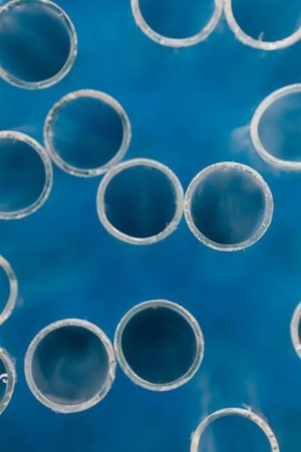
Utilizing soap bubbles as a model for cell membranes allows for a simplified‚ yet effective‚ visualization of complex biological structures. The bubble’s thin film directly represents the phospholipid bilayer‚ demonstrating how a hydrophobic core is sandwiched between hydrophilic heads – mirroring the cell membrane’s composition;
The bubble’s ability to stretch‚ contract‚ and even repair small tears directly parallels the fluidity and self-sealing capabilities of real cell membranes. Manipulating the bubble with a thread simulates the dynamic nature of membrane proteins and their influence on membrane shape.
Detailed observations and subsequent analysis‚ often documented in a PDF lab report‚ reinforce these concepts‚ providing a concrete understanding of abstract biological principles.
Materials Used in the Bubble Lab
Essential materials include dish soap‚ corn syrup‚ water‚ thread‚ and a bubble wand‚ enabling students to construct and manipulate bubbles mimicking cell membrane properties.
Lab reports (often PDF format) detail precise measurements and observations of these materials’ impact on bubble formation and stability.
Dish Soap as a Phospholipid Bilayer
Dish soap molecules effectively model the phospholipid bilayer structure of a cell membrane due to their amphipathic nature – possessing both hydrophilic (water-attracting) and hydrophobic (water-repelling) regions.
Similar to phospholipids‚ soap molecules arrange themselves with the hydrophilic heads facing outwards towards the water and the hydrophobic tails clustering inwards‚ creating a bilayer.
This bilayer forms the bubble’s skin‚ demonstrating the basic structural component of cell membranes. Lab reports‚ frequently provided as PDF documents‚ emphasize this analogy.
Students analyze how the soap film’s flexibility and self-sealing capabilities mirror the dynamic properties of a real cell membrane‚ crucial for maintaining cellular integrity.
The lab’s success hinges on understanding this fundamental relationship‚ often assessed through questions within the PDF analysis‚ requiring students to connect the model to biological reality.
Corn Syrup and Water: Viscosity and Membrane Fluidity
The combination of corn syrup and water in the bubble solution directly influences the membrane’s fluidity‚ mirroring the effect of cholesterol in real cell membranes. Corn syrup increases the viscosity of the solution‚ slowing down movement.
This increased viscosity represents a less fluid membrane‚ while a higher water concentration promotes greater fluidity. Students observe how solution composition impacts bubble stability.
Lab reports‚ often distributed as PDF files‚ require students to correlate viscosity changes with the bubble’s ability to stretch and repair itself.
Analyzing the bubble’s response to manipulation helps illustrate how membrane fluidity affects essential cellular processes like transport and signaling.
Understanding this relationship is key to grasping the dynamic nature of cell membranes‚ a core concept frequently tested in the PDF-based lab assessment.
Thread and Bubble Wand: Manipulating the Membrane
The bubble wand serves as a tool to create the initial membrane structure‚ while the thread simulates external forces acting upon the cell membrane. Carefully inserting the thread allows students to observe the bubble’s ability to reseal.
This demonstrates the self-sealing capabilities of the phospholipid bilayer‚ a crucial characteristic of living cell membranes. The success of splitting the bubble highlights membrane integrity.
Lab reports‚ commonly provided as a PDF document‚ often ask students to analyze why some bubbles split cleanly while others rupture. This relates to bilayer strength.
Students document observations about the bubble’s response to the thread‚ connecting it to the membrane’s fluidity and the presence of repair mechanisms.
The thread manipulation is a key component assessed in the PDF lab analysis‚ testing comprehension of membrane dynamics and structural resilience.
Procedure of the Cell Membrane Bubble Lab
The lab involves creating a bubble solution‚ carefully forming bubbles‚ and then using thread to split them‚ observing membrane behavior for PDF analysis.
Creating the Bubble Solution
The foundation of this lab lies in meticulously crafting the bubble solution‚ a direct analog to the cellular environment. Begin by combining dish soap – representing the phospholipid bilayer – with both corn syrup and water. The precise ratios are crucial; corn syrup adds viscosity‚ mimicking membrane fluidity‚ while water acts as the solvent.
Gentle mixing is essential to avoid excessive foam‚ which can hinder observation. Students often record the exact measurements used in their lab notebooks‚ later included in a comprehensive PDF report detailing the experimental setup. This solution’s properties directly influence bubble formation and stability‚ impacting the demonstration of cell membrane characteristics. Careful preparation ensures a successful and insightful experiment‚ leading to accurate data for analysis and a well-documented PDF submission.
Forming and Observing the Bubble
Carefully dip the bubble wand into the prepared solution‚ creating a thin film. Gently withdraw the wand‚ allowing a bubble to form. Observe its iridescent surface‚ noting its fluidity and how it responds to slight disturbances. Students should meticulously document these initial observations in their lab reports‚ often including photographs for a visual record.
Pay close attention to the bubble’s structural integrity and how it maintains its shape‚ mirroring the cell membrane’s ability to contain cellular contents. Detailed descriptions of bubble behavior are vital for the PDF analysis‚ connecting observations to cellular functions. Record any imperfections or variations in the bubble’s surface‚ as these can relate to membrane permeability and repair mechanisms‚ ultimately contributing to a complete PDF submission.
Splitting the Bubble with Thread
Slowly insert the thread into the bubble solution‚ then carefully lift it through the bubble’s film. Observe as the bubble splits into two separate bubbles‚ demonstrating the membrane’s ability to divide and reseal. This action models cellular processes like vesicle formation and cell division.
Document the splitting process‚ noting the ease or difficulty of separation and the resulting bubble shapes. This observation is crucial for the PDF lab report‚ illustrating membrane flexibility and self-repair capabilities. Analyze how the thread disrupts the bilayer‚ and how the bubble attempts to restore its integrity. Detailed descriptions and diagrams are essential for a comprehensive PDF analysis‚ connecting this demonstration to real cell membrane dynamics.
Cell Membrane Concepts Demonstrated
This lab vividly illustrates selective permeability‚ phospholipid bilayer structure‚ and membrane fluidity‚ concepts often detailed in a comprehensive cell membrane bubble lab answers PDF.
Selective Permeability and Bubble Integrity
The bubble’s skin‚ much like a cell membrane‚ isn’t freely permeable; it resists certain disruptions while yielding to others‚ demonstrating selective permeability. Observing how easily the bubble is broken‚ or maintains its shape‚ parallels a cell’s response to external factors.
This concept is crucial for understanding how cells regulate what enters and exits‚ a key topic often explored in a cell membrane bubble lab answers PDF. The bubble’s integrity‚ its ability to hold together‚ reflects the cell membrane’s role in maintaining cellular homeostasis.
Any tear or puncture in the bubble‚ similar to damage in a cell membrane‚ compromises its structure and function‚ leading to collapse. Detailed analysis‚ often found within a lab report PDF‚ highlights this relationship.
Phospholipid Bilayer Structure
The soap bubble itself serves as a compelling model for the phospholipid bilayer‚ the fundamental structure of cell membranes. Soap molecules possess both hydrophilic (water-attracting) and hydrophobic (water-repelling) properties‚ mirroring the amphipathic nature of phospholipids.
This dual nature causes the soap molecules to arrange themselves with their heads facing outwards (towards water) and tails inwards‚ creating a bilayer – just like in a cell membrane. Understanding this structure is often a core component of a cell membrane bubble lab answers PDF.
The bubble’s thin‚ flexible skin visually represents the bilayer’s fluidity and its ability to self-seal‚ crucial for maintaining cell integrity. Detailed diagrams and explanations are frequently included in lab report PDF resources.
Membrane Fluidity and Repair
The bubble’s dynamic nature beautifully illustrates membrane fluidity – the ability of the lipid bilayer to move and change shape. Small punctures or tears in the bubble quickly reseal‚ demonstrating the membrane’s self-repair capabilities‚ a vital function of cell membranes.
This self-sealing is due to the hydrophobic interactions between the lipid tails‚ which spontaneously rearrange to minimize contact with water. Observing this repair process is a key objective of the lab‚ often requiring detailed documentation in a cell membrane bubble lab answers PDF.
Students analyze how the bubble responds to disturbances‚ relating this to the cell’s ability to maintain its structural integrity despite external stressors. Lab reports frequently include explanations of these observations‚ often found within a comprehensive PDF guide.
Analyzing the Results of the Lab
Careful observation and data collection are crucial; students relate bubble behavior to cell membrane function‚ often documented and assessed in a PDF report.
Observations and Data Collection
Meticulous recording of observations is paramount during the cell membrane bubble lab. Students should document bubble formation‚ stability‚ and response to the thread manipulation.
Detailed notes on bubble size‚ shape‚ and lifespan before popping are essential. Observing how the bubble splits with the thread‚ and noting any irregularities‚ provides valuable data.
Quantitative data‚ such as approximate bubble diameter or time until rupture‚ can be collected. These observations‚ often compiled into a lab report – frequently a PDF document – form the basis for analysis.
Qualitative descriptions of bubble behavior‚ like the appearance of the film or any visible repairs‚ are also important. This comprehensive data collection allows for meaningful connections to cell membrane properties.
Relating Bubble Behavior to Cell Membrane Function
The bubble’s fragility mirrors the cell membrane’s susceptibility to disruption‚ highlighting the importance of its structure. Observing bubble repair after minor tears demonstrates the membrane’s self-sealing capabilities.
The thread splitting the bubble illustrates membrane fission during cell division or vesicle formation. Bubble solution viscosity relates to membrane fluidity‚ impacting permeability and function.
Analyzing these parallels allows students to grasp abstract cell membrane concepts in a tangible way. This connection is often a key component of the cell membrane bubble lab answers PDF‚ requiring students to articulate these relationships.
Understanding how bubbles maintain their shape and integrity provides insight into the cell’s ability to maintain homeostasis and compartmentalization.
Common Lab Report Questions & Answers (PDF Focus)
Frequently‚ lab reports ask students to explain how the bubble’s phospholipid bilayer relates to the cell membrane’s structure‚ often requiring diagrams. Another common question explores selective permeability‚ asking what substances could/couldn’t pass through.
The cell membrane bubble lab answers PDF often includes prompts about membrane fluidity and how viscosity impacts this property. Students are also asked to analyze bubble splitting‚ connecting it to cell division.
Expect questions about the limitations of the model – bubbles aren’t perfect representations of cells! Answers should demonstrate understanding of the model’s strengths and weaknesses.
Detailed PDFs typically provide scoring rubrics‚ emphasizing clear explanations and connections between observations and cell membrane functions.
Troubleshooting Common Issues
Premature popping often results from dirty wands or solutions; careful preparation is key. Splitting difficulty may require slower thread movement and practice.
Bubble Popping Prematurely
Frequent bubble failures during the cell membrane bubble lab often stem from contaminants. Ensure all equipment‚ especially the bubble wand and mixing containers‚ are scrupulously clean and free of residues. Even trace amounts of oils or other substances can disrupt surface tension.
Additionally‚ the bubble solution itself might be the culprit. Verify the correct proportions of dish soap‚ corn syrup‚ and water are used‚ as imbalances affect stability. Humidity also plays a role; drier air accelerates evaporation‚ leading to quicker popping. Finally‚ gentle handling is crucial – avoid drafts or accidental contact that could compromise the bubble’s delicate structure. Reviewing lab reports (often in PDF format) can highlight common errors.
Difficulty Splitting the Bubble
Challenges in splitting the bubble with thread often indicate issues with solution viscosity or thread technique. Ensure the bubble film is sufficiently strong and elastic – adjust corn syrup concentration if needed‚ increasing it slightly for greater resilience. Slowly and steadily lift the thread through the bubble‚ avoiding quick movements that can cause it to pop instead of divide.
A clean‚ unwaxed thread is essential; coatings can prevent smooth passage. Practice the motion to develop a consistent technique. If the bubble still resists splitting‚ consider the bubble’s size – larger bubbles are generally easier to manipulate. Detailed instructions and troubleshooting tips are frequently found within comprehensive cell membrane bubble lab reports‚ often available as a PDF resource.